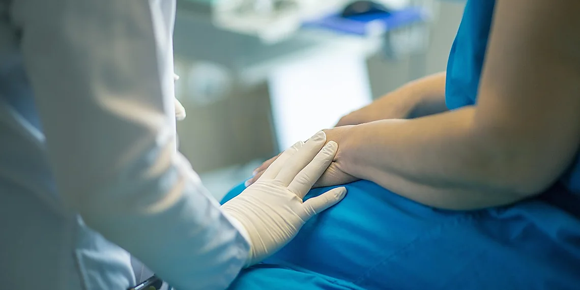

Imagine a world where medical diagnoses are faster, more precise, and accessible to everyone – that’s the promise of artificial intelligence in healthcare. But realizing that vision faces a significant hurdle: accurately labeling medical images is incredibly time-consuming and costly, often requiring teams of highly specialized experts meticulously annotating every detail.
The bottleneck isn’t just about labor hours; it’s also about data. Training robust AI models demands massive datasets, but acquiring these annotated datasets in the medical field is a monumental challenge due to privacy concerns and the sheer complexity of biological structures.
Now, what if we could leapfrog this limitation? Researchers are pioneering a revolutionary approach leveraging quantum-inspired algorithms to dramatically reduce the amount of data needed for training. This breakthrough allows us to achieve exceptional results even with limited examples – a game changer particularly relevant for tasks like breast cell segmentation, where subtle variations can be critical.
This article delves into how these novel techniques are transforming clinical AI, offering a glimpse at a future where diagnostic tools are smarter, more efficient, and ultimately, improve patient outcomes.
The Annotation Bottleneck in Medical AI
Developing effective Artificial Intelligence models for medical image analysis hinges on having high-quality, meticulously labeled datasets. However, creating these datasets presents a significant bottleneck – the annotation process itself is incredibly demanding and time-consuming. Pathologists and other specialists are required to manually outline regions of interest in images, essentially teaching the AI what it’s looking for. This isn’t a simple task; medical images often exhibit complex textures, subtle variations, and overlapping structures that require deep expertise to interpret accurately.
The challenges extend beyond just visual complexity. ‘Inter-annotator variability,’ where different specialists might label the same region slightly differently, introduces another layer of difficulty. Subtle differences in interpretation are common, particularly when dealing with nuanced features like cellular boundaries. This inconsistency needs to be accounted for during training and can significantly impact model performance. The abstract highlights a case where inter-annotator variations reach +/- 3 pixels – a seemingly small difference that can have profound consequences.
Consider the sheer volume of effort involved. As the recent arXiv paper (2512.02302v1) details, annotating even relatively modest mammary epithelial nuclei datasets can require pathologists to invest hundreds of hours of their valuable time. This represents a substantial cost, both in terms of financial resources and specialist availability. It also limits the scale of datasets that are realistically possible, potentially hindering the development of more robust and accurate AI solutions.
Ultimately, overcoming this annotation bottleneck is crucial for advancing medical AI applications. The research described in the abstract addresses this directly by demonstrating a model capable of achieving impressive results (95.5% Dice score) with a comparatively small dataset (599 images). This showcases the potential of innovative approaches – like the quantum-inspired edge enhancement used here – to mitigate the reliance on massive, laboriously annotated datasets and accelerate progress in fields such as breast cell segmentation.
Why Medical Data is So Hard to Label

Creating high-quality datasets for training medical AI models is a significant hurdle, particularly in areas like breast cancer diagnosis. Pathologists, who possess the necessary expertise to accurately label these images, face considerable challenges stemming directly from the nature of the data itself. Mammary epithelial nuclei images, for example, are incredibly complex, exhibiting subtle variations in appearance and often containing overlapping cells making precise delineation difficult.
A major compounding factor is inter-annotator variability. Even among experienced pathologists, there’s inherent subjectivity in interpreting these images. Differences in perception and judgment can lead to significant discrepancies in how they delineate cell boundaries – the study cited highlights this variation reaching plus or minus three pixels, a crucial detail when training AI for accurate segmentation. This inconsistency necessitates extensive review and reconciliation of annotations.
The sheer volume of effort required is staggering. Preparing even relatively small datasets often involves hundreds of hours of pathologist time dedicated solely to labeling. Consider that many images might contain minimal breast tissue (as noted in the study, only 4% of pixels represent breast tissue in some cases), or lack any relevant regions entirely – this further increases the workload and cost associated with dataset creation.
Quantum-Inspired Enhancement: Seeing the Edges
Accurate breast cell segmentation is a cornerstone of early cancer detection and diagnosis, but it’s notoriously difficult. Pathologists spend countless hours meticulously outlining cells in medical images, a process ripe for human error and inconsistency. A significant hurdle lies in the often-blurry or indistinct boundaries between individual cells – variations can easily reach plus or minus three pixels, making precise delineation challenging even among experienced professionals. New research published on arXiv is tackling this head-on with an innovative approach: quantum-inspired edge enhancement.
The key to their breakthrough lies in a novel application of what they call ‘quantum-inspired Gabor filters.’ Now, don’t worry about the ‘quantum’ part – it’s more about using concepts from quantum mechanics to design these filters. Think of Gabor filters as specialized magnifying glasses that highlight specific edges and features within an image at different scales. Just like a regular magnifying glass helps you see tiny details, these filters sharpen cell boundaries, making them clearer and more distinct. By applying multiple filters at varying sizes, the system can capture a wider range of edge characteristics.
The real benefit is how this enhanced edge information feeds into the segmentation process. The team essentially created an extra ‘channel’ of data – a fourth input for their AI model – representing these sharpened boundaries. This allows the algorithm to better distinguish between individual cells, even when those boundaries are vague or inconsistent across different image annotations. This is particularly crucial in scenarios where images contain very little breast tissue or have large areas with no breast regions at all, as this new technique helps the model focus on what *is* present.
Remarkably, using just 599 training images, the framework achieved an impressive Dice score of 95.5% – a measure of how well the predicted segmentation aligns with the ground truth. This demonstrates that even with limited data and significant boundary variation challenges, this quantum-inspired edge enhancement technique represents a substantial leap forward in automating and improving breast cell segmentation, ultimately promising to reduce pathologist workload and improve diagnostic accuracy.
How Multi-Scale Gabor Filters Work (Simplified)
Imagine trying to draw a line around something that isn’t perfectly clear – like outlining a cell under a microscope. Pathologists often disagree slightly on where those lines should be, leading to inconsistencies in how images are labeled. This makes training AI models tricky; they learn from these labels and if the ‘truth’ is blurry, the AI’s understanding will be too. The research described here tackles this problem directly by sharpening the edges of cells before the AI even sees them.
One way to sharpen those edges is through something called Gabor filters. Think of them as tiny magnifying glasses that look for specific patterns – in this case, changes in brightness and contrast that define a cell’s boundary. These filters aren’t just one size; they come in different scales, like looking at the same object with a wide-angle lens versus a telephoto lens. This multi-scale approach helps capture edges even when cells vary significantly in size or shape.
The team used a ‘quantum-inspired’ technique to improve these Gabor filters, essentially making them more effective at highlighting those crucial boundaries. The result is an extra layer of information—a fourth input channel—that the AI can use. This enhanced edge detection dramatically reduces the impact of differences in how pathologists draw their lines, allowing the AI to learn a more consistent and accurate representation of breast cells.
Stabilizing the AI with Adaptive Loss & Sampling
A major hurdle in training effective AI models for breast cell segmentation is the severe class imbalance inherent in medical image datasets. Typically, only a tiny fraction – around 4% in our study – of pixels actually represent the target cells (mammary epithelial nuclei). The vast majority are background noise. This disparity can lead to models that prioritize learning the background, effectively ignoring or misclassifying the few precious breast cells. To combat this, we’ve developed an adaptive Dice loss function designed specifically for this challenge. Unlike standard Dice loss, our approach dynamically adjusts its weighting based on the current performance of the model on different classes – in this case, foreground (breast cells) and background.
The core innovation lies within three key components. First, the adaptive Dice loss continually re-evaluates the relative importance of each class during training, focusing more attention on areas where the model is struggling to accurately identify breast cells. Second, we integrated boundary-aware terms into the loss function. Given that inter-annotator variability in outlining cell boundaries can be significant (reaching +/- 3 pixels), these terms penalize errors specifically at cell edges, encouraging the model to learn finer distinctions and improve segmentation accuracy. Finally, an automatic positive weighting scheme ensures the foreground class receives consistent importance, preventing it from being overwhelmed by the background.
Beyond the loss function, we also implemented a strategic sampling approach that prioritizes difficult examples. Because 60% of our images contained no breast regions at all (effectively ’empty’ images), simply feeding these into training would be counterproductive. Our sampling strategy intelligently selects images and within those images, segments, that present the greatest challenge to the model – for instance, cases with poorly defined cell boundaries or unusual tissue morphology. This allows the model to learn from its mistakes and improve its ability to handle complex scenarios, ultimately boosting overall performance.
By combining this adaptive loss function with intelligent sampling, we’ve created a framework capable of achieving remarkable results with limited training data – a 95.5% Dice score using just 599 images. This represents a significant step forward in automating the laborious process of medical image annotation and promises to accelerate research and improve diagnostic accuracy in breast cancer detection.
Taming Class Imbalance: The Adaptive Loss Function
Medical image analysis frequently suffers from a problem known as class imbalance. In the context of breast cell segmentation, this means that the vast majority of pixels in an image represent background – for example, tissue surrounding breast cells – while only a small percentage actually belong to the target object (the breast cells themselves). This disparity can severely bias machine learning models, causing them to prioritize predicting the common class (background) and perform poorly on identifying the rarer, but crucial, breast cells. The dataset described in arXiv:2512.02302v1 highlights this challenge, noting that only 4% of pixels represent breast tissue and many images contain no breast regions at all.
To combat this class imbalance, the researchers developed an adaptive Dice loss function as a core component of their framework. The standard Dice loss can be overwhelmed by the dominant background class; the adaptive version dynamically adjusts its weighting to give more emphasis to the underrepresented breast cells. This is further enhanced with boundary-aware terms that specifically penalize errors at cell boundaries, which are often areas of significant inter-annotator variability (reaching +/- 3 pixels in this study). These boundary terms help refine segmentation accuracy and improve delineation.
Finally, an automatic weighting strategy ensures that the loss function prioritizes difficult examples. Images or regions within images where the model is struggling to accurately segment breast cells receive higher weights during training. This targeted approach focuses learning on the most challenging cases, leading to a more robust and accurate segmentation model even with limited training data – achieving a 95.5% Dice score from only 599 training images.
Results & The Future of Limited-Data AI
The research presented in arXiv:2512.02302v1 demonstrates a remarkable breakthrough in breast cell segmentation, achieving an impressive 95.5% Dice score with a remarkably small training dataset of just 599 images. This is particularly significant given the inherent challenges of mammary epithelial nuclei datasets – they often feature extremely low tissue representation (only 4% of pixels) and a substantial proportion of images lacking any breast regions at all. The ability to attain such high accuracy with so little data directly addresses a major bottleneck in medical image analysis: the laborious and expensive process of manual annotation, which traditionally requires hundreds of hours from skilled pathologists.
The success isn’t solely attributable to clever architecture; it’s driven by innovative techniques designed to maximize information extraction. The researchers employed quantum-inspired edge enhancement using multi-scale Gabor filters, effectively creating a fourth input channel that sharpens boundaries – a critical factor given inter-annotator variations can reach +/- 3 pixels. Furthermore, a stabilized multi-component loss function incorporating adaptive Dice loss and boundary-aware terms alongside automatic positive weighting further refined the model’s performance. This combination of techniques allows for significantly improved segmentation quality even when dealing with sparse and noisy data, representing a substantial advancement over conventional approaches.
The implications extend far beyond simply improving breast cell segmentation accuracy. This methodology offers a pathway to developing clinical AI tools that require dramatically less labeled data – reducing the burden on medical professionals and potentially accelerating the development of diagnostic solutions for other diseases. Imagine applying similar techniques to segmenting tumors in lung scans or identifying anomalies in retinal images, all while minimizing the need for extensive manual annotation. The reduced data requirements also open doors for broader accessibility; institutions with limited resources can now explore AI-powered diagnostics more readily.
Looking ahead, this work paves the way for a future where clinical AI development is less reliant on massive datasets and more focused on intelligent feature engineering and quantum-inspired enhancements. The team’s approach highlights the potential of leveraging unconventional techniques to overcome data scarcity challenges in medical imaging – ultimately leading to faster diagnoses, reduced workloads for pathologists, and potentially earlier detection of critical conditions.
Beyond the Numbers: What This Means for Clinical Practice
The breakthrough demonstrated in this research has significant potential to ease the workload of pathologists. Currently, accurately identifying and segmenting breast cells within medical images is a painstakingly manual process, demanding hundreds of hours of expert annotation. This new approach, achieving a 95.5% Dice score with only 599 training images, drastically reduces the data requirements for reliable segmentation. This means less time spent on tedious labeling tasks, freeing up pathologists to focus on more complex diagnostic evaluations and patient care.
Beyond simply reducing workload, this technology promises to accelerate the diagnostic process. Faster and more accurate breast cell segmentation can lead to quicker diagnoses, potentially enabling earlier detection of abnormalities and ultimately improving patient outcomes. The quantum-inspired edge enhancement, which addresses inherent challenges in image clarity and inter-annotator variability, plays a crucial role in achieving this level of precision even with limited data. This contributes to a more efficient and reliable diagnostic workflow.
Looking ahead, the framework’s success in breast cell segmentation suggests broader applicability within medical imaging. The ability to train accurate AI models using limited datasets is particularly valuable for rare diseases or conditions where large annotated image collections are simply not available. Researchers envision extending this approach – combining quantum-inspired enhancement with adaptive loss functions – to other areas like lung nodule detection, cardiac MRI analysis, and even neurological image interpretation, paving the way for more accessible and powerful clinical AI tools.
The convergence of quantum computing principles and artificial intelligence is proving transformative, particularly within the realm of medical imaging. This research unequivocally demonstrates a powerful new approach to tackling complex challenges like accurate breast cell segmentation, showcasing how we can achieve exceptional results even when faced with constrained datasets. The implications extend far beyond this specific application; it signals a broader paradigm shift in AI development, one where efficiency and precision are prioritized alongside sheer computational power. By leveraging quantum-inspired algorithms, we’re not just improving existing technologies but fundamentally rethinking how medical AI is built and deployed, potentially democratizing access to advanced diagnostics worldwide. This breakthrough holds the promise of faster, more reliable diagnoses, reduced patient anxiety, and ultimately, improved health outcomes for countless individuals. The future of healthcare will undoubtedly be shaped by innovations like this, pushing the boundaries of what’s possible in disease detection and treatment. To truly grasp the scope of this revolution and its potential to reshape medical practice, we urge you to delve deeper into the fascinating world of quantum-inspired AI and explore how it’s poised to revolutionize healthcare as we know it.
Learn more about the burgeoning field of quantum-inspired AI – resources are readily available online and through leading academic institutions. Consider exploring introductory courses or research papers to understand its core principles and potential applications beyond medical imaging. The possibilities are vast, and your engagement can contribute to a future where healthcare is smarter, faster, and more accessible for everyone.
Continue reading on ByteTrending:
Discover more tech insights on ByteTrending ByteTrending.
Discover more from ByteTrending
Subscribe to get the latest posts sent to your email.